
The roof, or superior wall, of the orbit separates the orbit from the anterior cranial fossa.

The optic foramen provides passage for the optic nerve (CN II) and ophthalmic artery to exit the skull. It is medial to the superior orbital fissure. The apex of the orbit is the optic foramen (canal), bound medially by the body of the sphenoid bone and laterally by the lesser wing of the sphenoid bone.

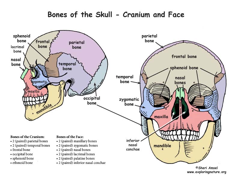
Finally, a few words about the most common pathological conditions related to the orbit will be discussed. This article will discuss the bones of the orbit, their articulations, and the most important anatomical landmarks. Orbital fractures, inflammatory and neoplastic processes Optic foramen (canal), lacrimal fossa, lacrimal groove, anterior and posterior ethmoidal foramina, trochlea, superior and inferior orbital fissuresĮyeballs, orbital fascia, orbital fat, extraocular muscles, neurovasculature, lacrimal apparatus Medial: orbital plate of the ethmoid bone, lacrimal bone, frontal process of the maxilla, lesser wing of the sphenoid boneįloor (inferior): orbital surface of the maxilla, zygomatic bone, palatine boneįrontomaxillary, zygomaticomaxillary, frontozygomatic (or zygomaticofrontal), fronto-ethmoidal, sphenofrontal (or frontosphenoidal), sphenozygomatic, frontolacrimal, spheno-ethmoidal, lacrimomaxillary Roof (superior): orbital part of the frontal bone, lesser wing of the sphenoid bone Lateral margin: zygomatic process of the frontal bone and the zygomatic bone and its frontal process Infra-orbital margin: zygomatic process of the maxilla and the zygomatic bone Medial margin: frontal process of the maxilla Many Friendly Zebras Enjoy Lazy Summer Picnic Maxilla, Frontal bone, Zygomatic bone, Ethmoid bone, Lacrimal bone, Sphenoid bone, and Palatine bone The bones that make up the orbit contain several foramina and fissures through which important neurovascular structures (such as the optic nerve (CN II)) pass through on their way from the brain to the eye and face and vice versa. The cavity surrounds and provides mechanical protection for the eye and soft tissue structures related to it. By definition, the orbit (bony orbit or orbital cavity) is a skeletal cavity comprised of seven bones situated within the skull.


 0 kommentar(er)
